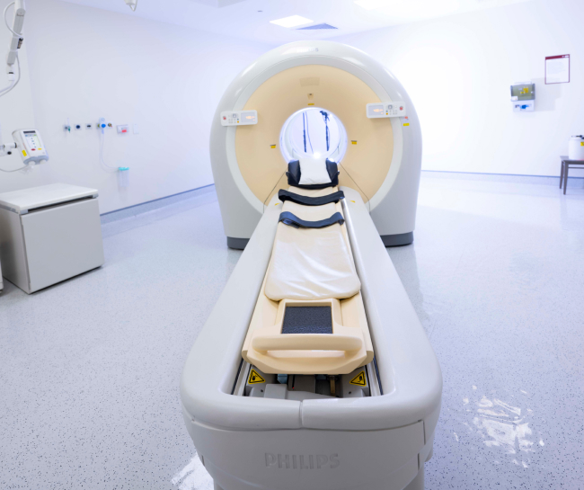
SPECT-CT and PET-CT
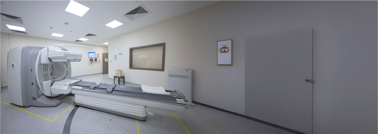
ر SPECT-CT وPET-CT في معهد برجيل للسرطان أدوات تشخيصية متقدمة للكشف عن أنواع السرطان المختلفة وتحديد مراحلها ومراقبتها. تجمع أنظمة التصوير الهجين هذه بين الطب النووي والتصوير المقطعي المحوسب
Key Features
Both SPECT-CT and PET-CT offer several features that make them invaluable in cancer diagnostics and management:
Cancers Diagnosed and Monitored with SPECT-CT and PET-CT
These imaging systems are used to diagnose, stage, and monitor a wide range of cancers, including:
- Lung cancer
- Breast cancer
- Lymphoma (Hodgkin’s and non-Hodgkin’s)
- Colorectal cancer
- Prostate cancer
- Thyroid cancer
- Melanoma
- Head and neck cancer
- Bone metastases (secondary cancer spread to bones)
- Neuroendocrine tumors
- Other solid tumors and metastatic cancers
Benefits of SPECT-CT and PET-CT
These advanced imaging systems provide numerous benefits to patients and physicians:
Accurate Diagnosis
PET-CT and SPECT-CT offer a comprehensive view of cancer activity and anatomical structures, allowing for more accurate diagnosis and staging of cancer.
Early Detection
PET-CT can detect metabolic changes in cells before structural changes are visible, allowing for earlier diagnosis and improved treatment outcomes.
Treatment Planning
The detailed images provided by these scans guide radiation oncologists and surgeons in planning precise treatments, such as radiation therapy or surgical tumor removal.
Monitoring Treatment Response
Both technologies are invaluable for assessing how well a treatment is working by monitoring changes in cancerous activity, helping to adjust treatment plans if necessary.
Minimally Invasive
Both imaging techniques are non-invasive and painless, offering patients a safe way to undergo comprehensive cancer diagnostics.
Our Approach to Imaging and Diagnosis
At Burjeel Cancer Institute, SPECT-CT and PET-CT are integral to our comprehensive approach to cancer care:
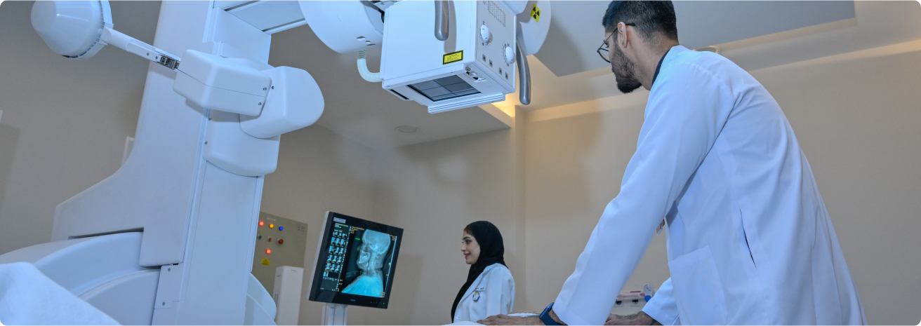
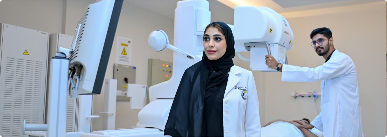
Multidisciplinary Collaboration
Our radiologists, nuclear medicine physicians, oncologists, and surgeonscollaborate to interpret imaging results and develop personalized treatment plans.
Personalized Diagnostic Plans
Every patient’s imaging and diagnostic needs are tailored based on their type of cancer, medical history, and treatment goals.
Advanced Imaging Techniques
We utilize the latest PET-CT and SPECT-CT technology to ensure the highest level of accuracy in cancer diagnosis and treatment planning.
Focus on Precision
These imaging technologies provide precise details about the location, size, and spread of tumors, allowing for targeted and effective treatment.
Patient Journey
Patients undergoing surgery with G-Arm and O-Arm imaging at Burjeel Cancer Institute can expect a well-coordinated and supportive experience:
Initial Consultation
A comprehensive evaluation with the bladder cancer care team, including diagnostic imaging and tests to assess the extent of the disease.
Personalized Treatment Plan
Based on the patient’s diagnosis, preferences, and overall health, a customized treatment plan is developed.
Treatment and Support
Patients undergo the recommended treatments, supported by a multidisciplinary team and personalized supportive care services.
Follow-Up Care
After treatment, patients receive regular follow-ups to monitor recovery, assess treatment response, and manage long-term health concerns.
